Cervical Disc Herniation
Cervical Disc Herniation
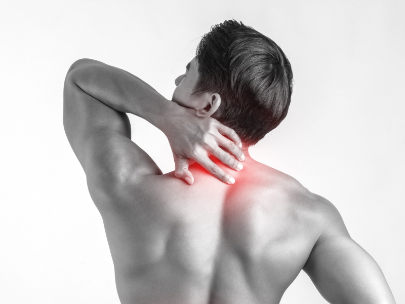
What Is a Cervical Disc Hernia?
The spine consists of a series of interlinked bones, called vertebra. The vertebras surround the spinal cord and protect it from injury. The nerves branch from the spinal cord and travel to other parts of the body, thereby forming the link between the brain and the rest of the body.
The brain moves the muscles by a message carried by the nerves. The nerves also carry senses such as pain and heat from the body to the brain.
The vertebral bodies are connected to each other with one disc and two small joints, named facet joints. The disc is the most important structure that connects one disc to another. It is formed by a strong connective tissue, and acts as a cushion or shock absorber between the vertebras. The discs and facet joints enable movement of the vertebra, thereby making it possible to bend your neck or back, or to rotate them.
The disc is composed of a strong outer layer named "annulus fibrosus", and a jelly-like core named "nucleus pulposus".
With increasing age, the center of the disc begins to lose water content, and the disc cannot perform its shock absorbing function as much as it used to. The outer layer may also rupture as the disc continues to degenerate. This may cause overflowing (disc herniation or disc rupture) of the disc through a tear in the outer layer, and into the space that harbors the nerves and the spinal cord.
The herniated disc may then press against the nerves and cause pain, loss of sensation, tingling, and weakness on the arms and shoulders. Your doctor may detect changes in the strength of your muscles, reflexes, or senses. In rare instances, the herniated dics may cause leg problems by comressing the spinal cord.
How Is It Diagnosed?
A clinical examination focused on finding the source and type of pain, and also a careful examination of any muscle weakness, sensory loss, and abnormal reflex are often enough to diagnose and locate discal hernia.
Your doctor's diagnosis will be confirmed with X-rays, computerized tomography, and magnetic resonance imaging. X-rays can show bony projections and narrowing of the disc spaces that occur with wear and degeneration of the spine, however it cannot show disc hernation or the nerves branching from the spinal cord. CT and MRI (the gold standard) enable detailed images of all structures of the spine (vertebrae, discs, spinal cord and nerves), and detect most disc herniations. In addition, by conducting nerve studies, nerve injury—possibly caused by a disc hernia—can be investigated.
What Treatments Are Available?
Most patients with cervical disc hernia can improve without any treatment. In patients with persistent pain, there are a number of choices. Many drugs and non-surgical modalities will help decrease pain related to cervical disc hernia. Surgical treatment will be required for some patients.
Non-surgical Treatment in Cervical Disc Herniation
Most patients will improve with non-surgical (medical) treatment or conservative care.
Your doctor may recommend various treatments, such as a short period of bed rest, short-term use of a cervical collar, anti-inflammatory drugs to decrease nerve irritation, analgesics for pain control, physical therapy, exercise or epidural steroid injections. The aim of nonsurgical treatments is to decrease nerve irritation caused by the herniated disc, relieve the pain, and to improve the general condition of the patient. These results can be attained in most patients with disc hernia using a well-organized treatment program that integrates numerous treatment methods. Ask your doctor if you will be able to continue to work during treatment.
Following the onset of pain due to cervical disc hernia, a brief (1-2 days) period of rest may be useful. Returning to activities after this brief period of rest is crucial to prevent joint stiffness and muscle weakness. With the assistance of a nurse or physiotherapist, your doctor may teach you the specific exercises that will strengthen your neck. These exercises may be practiced at home, however you may need to visit a physiotherapist for a more special program that suits your abilities. The exercises must be performed exactly as your doctor or physiotherapist has shown. Your doctor or physiotherapist may decrease the pain, inflammation, and muscle spasm by applying treatments such as traction, heat or cold application, and manual (massage) therapy.
Drugs and Pain Treatment
Drugs that are used to control pain are called painkillers (analgesics). In most situations, the pain responds to commonly used over-the-counter medications like aspirin, ibuprophen, naproxen, and acetaminophen. If you have severe or persistent pain, your doctor may prescribe narcotic analgesics for a short time. Muscle relaxants may be added in some cases. A higher dose will not accelerate your healing. Possible side effects include: nausea, constipation, dizziness, and vertigo, with a chance of addiction. All drugs must be used exactly in the manner prescribed. Inform your doctor of all the drugs you are using (including over-the-counter drugs) and whether they have worked. Also, inform your doctor of any allergic reactions to drugs.
A group of analgesics named non-steroidal anti-inflammatory drugs (NSAIDs) may be added to control the inflammation in the nerves and the neighboring tissues. These include prescription drugs such as ibuprophen, naproxen, and diclofenac. If your doctor has given you analgesics or anti-inflammatory drugs, you have to be careful of side effects such as gastric irritation or bleeding. You should follow up with your doctor regarding possible long-term negative consequences of prescribed or non-prescribed analgesics and NSAIDs. In intense arm and neck pain, corticosteroid drugs (tablets or injections) are also prescribed occasionally for their strong anti-inflammatory effects. Corticosteroids may have side effects similar to NSAIDs. You should talk with your doctors about the benefits and risks of these drugs.
Epidural injections or "blocks" may be used to relieve intense arm pain. These are corticosteroid (cortizone) injections into the epidural space (the space around the nerves) by a doctor trained on this technique. The first injection may be supplemented with one or two subsequent injections. These are often done in an interactive rehabilitation and treatment program to decrease the inflammation of the disc and nerve.
Trigger point injections are local anesthetic (corticosteroids may be added in some cases) injections made directly into the painful soft tissues and muscles along the spine. Although they are helpful for pain control in some cases, they do not correct the herniated disc.
Surgical Treatment in Cervical Disc Herniation
Surgery may be necessary in patients who did not benefit from other therapies. The goal of surgery is removal of the part of the disc that is pressing on the nerve. This is done with a procedure named discectomy. The hernia is accessed through a skin incision on the front or back of the neck. The decision to perform surgery from the front (anterior approach) or the back (posterior approach) is affected by numerous factors such as the location of disc herniation, experience of the surgeon, and the patient’s preference. In both approaches, the part of the disc pressing on the nerve is removed, often with good results. In the front approach, most of the disc will need to be removed to reach the herniated disc, which will generally require a fusion.
The greatest disadvantage of fusion surgery is elimination of the movement at the fusion site. Discectomy at a single level does not pose a significant disadvantage with respect to neck mobility. This is because the lost mobility of this segment is compensated and tolerated by other intact levels. However, increased movement and workload imposed on the upper and lower segments will result in wear, neck hernia and pain in these areas. Today, because of developing technology, mobile prostheses can be inserted in the space created by disc removal instead of perfomring a fusion.
However, prostheses are not appropriate for every patient. Ideal candidates are patients who are relatively younger, whose facet joints have not degenerated, and have preserved disc heights. Your doctor will decide best whether a prosthesis is appropriate for you.
What Can I Expect Post-Surgery?
Most patients can go home shortly after surgery—sometimes less than 24 hours. Your doctor will inform you of when you can return to your normal daily activities after surgery.
A comprehensive postoperative rehabilitation program is essential to completely fulfill the activities of daily life. Most patients benefit from a postoperative exercise and physical therapy program under surveillance. Ask your doctor about exercises that will facilitate your recovery. Surgery is very effective in the alleviation of shoulder and arm pain due to herniated disc. However, some pain may continue after surgery.
Most patients respond well to discectomy, however as in all types of surgery, there are some risks. Bleeding, infection, or injury to the spinal cord or nerves are among the risks. Also, persistence or recurrence after surgery are also possible. In 3-5 percent of patients, the disc may rupture again and result in future symptoms.
